Assessment of acute pancreatitis using the CT severity index and modified CT severity index
Abstract
Background: To assess the severity of acute pancreatitis (AP) using computed tomography (CT) severity index (CTSI) and modified CT severity index (MCTSI), to correlate with clinical outcome measures, and to assess concordance with severity grading, as per the revised Atlanta classification (RAC).
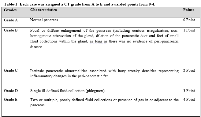
Material and Methods: This is a prospective study, conducted from August 2019 to July 2020, in the Department of Radiology, Al Ameen Medical College. A total of 70 patients referred from the Department of Medicine and Department of Surgery, presented with the chief complaint of epigastric pain, nausea and vomiting and CECT abdomen were suggestive of acute pancreatitis were included in this study. Assessment of severity of acute pancreatitis was done in all cases by Balthazar CTSI scoring and Mortele Modified CTSI scoring.
Results: In the present study total 70 cases of acute pancreatitis cases were included in the study. These patients underwent CT abdomen and pelvis, later images were reviewed by the radiologist. The maximum patients were in the age group of 21 to 40 years [n=33 (47.1%)]. Majority of the cases were categorized as mild pancreatitis according to Balthazar CTSI score. Majority of the cases were categorized as severe pancreatitis using the Modified Mortele CTS score. Whereas, organ failure, moderate and severe category in modified Mortele CTSI, mild, moderate, severe category in Balthazar CTSI.
Conclusion: In conclusion CECT was found to be an excellent imaging modality for diagnosis, establishing the extent of the disease process and in grading its severity.
Downloads
References
Spanier BW, Dijkgraaf MG, Bruno MJ. Epidemiology, aetiology and outcome of acute and chronic pancreatitis: An update. Best Pract Res Clin Gastroenterol. 2008;22(1):45-63. doi: 10.1016/j.bpg.2007.10.007.
Roberts SE, Akbari A, Thorne K, Atkinson M, Evans PA. The incidence of acute pancreatitis: impact of social deprivation, alcohol consumption, seasonal and demographic factors. Aliment Pharmacol Ther. 2013;38(5):539-548. doi: 10.1111/apt.12408.
Chatila AT, Bilal M, Guturu P. Evaluation and management of acute pancreatitis. World J Clin Cases. 2019;7(9):1006-1020. doi: 10.12998/wjcc.v7.i9.1006.
McKay C, Evans S, Sinclair M, Carter C, Imrie C. High early mortality rate from acute pancreatitis in Scotland, 1984-1995. Br J Surg. 1999;86(10):1302-1305. doi: 10.1046/j.1365-2168.1999.01246.x.
Secknus R, Mossner J. Changes in incidence and prevalence of acute and chronic pancreatitis in Germany. Der Chirurg. 2000;71(3):0249-0252.
Sharma V, Rana SS, Sharma RK, Kang M, Gupta R, Bhasin DK. A study of radiological scoring system evaluating extrapancreatic inflammation with conventional radiological and clinical scores in predicting outcomes in acute pancreatitis. Ann Gastroenterol. 2015;28(3):399 404.
Agarwal N. Assessment of severity in acute pancreatitis. Am J Gastroenterol. 1991;86(10):1385-1391.
Balthazar EJ, Freeny PC, vanSonnenberg E. Imaging and intervention in acute pancreatitis. Radiol. 1994;193(2):297-306. doi: 10.1148/radiology.193.2.7972730.
Balthazar EJ, Robinson DL, Megibow AJ, Ranson JHC. Acute pancreatitis: value of CT in establishing prognosis. Radiol. 1990;174(2):331-336. doi: 10.1148/radiology.174.2.2296641.
Mortele KJ, Mergo P, Taylor H, Ernst M, Ros PR. Renal and perirenal space involvement in acute pancreatitis: state-of-the-art spiral CT findings. Abdom Imaging. 2000;25(3):272-278. doi: 10.1007/s002610000032.
Ranson JHC, Rifkind KM, Roses DF, Fink SD, Eng K, Spencer FC. Prognostic signs and the role of operative management in acute pancreatitis. Surg Gynecol Obstet. 1974;139(1):69-81.
The Revised Atlanta Classification of Acute Pancreatitis: A Work Still in Progress? JOP. 2015;16(4):356 364.
Knaus WA, Draper EA, Wagner DP, Zimmerman JE. APACHE II: a severity of disease classification system. Crit Care Med. 1985;13(10):818-829.
Vincent JL, Moreno R, Takala J, Willatts S, De Mendonça A, Bruining H, et al. The SOFA (Sepsis related Organ Failure Assessment) score to describe organ dysfunction/ failure. On behalf of the Working Group on Sepsis Related Problems of the European Society of Intensive Care Medicine. Intensive Care Med. 1996;22(7):707 710. doi: 10.1007/BF01709751.
Steinberg W, Tenner S. Acute pancreatitis. N Engl J Med. 1994;330:1198-1210. doi: 10.1056/NEJM199404283301706.
Mendez Jr G, Isikoff MB, Hill MC. CT of acute pancreatitis: interim assessment. Am J Roentgenol. 1980;135(3):463-469. doi: 10.2214/ajr.135.3.463.
Johnson CD, Abu Hilal M. Persistent organ failure during the first week as a marker of fatal outcome in acute pancreatitis. Gut. 2004;53(9):1340 1344. doi: 10.1136/gut.2004.039883.
Banday IA, Gattoo I, Khan AM, Javeed J, Gupta G, Latief M. Modified computed tomography severity index for evaluation of acute pancreatitis and its correlation with clinical outcome: a tertiary care hospital based observational study. JCDR. 2015;9(8):TC01-5. doi: 10.7860/JCDR/2015/14824.6368.
Balthazar EJ. Acute pancreatitis: assessment of severity with clinical and CT evaluation. Radiol. 2002;223(3):603-613. doi: 10.1148/radiol.2233010680.
Melkundi S, Anand N. Modified computed tomography severity index in acute pancreatitis. J Evol Med Dental Sci. 2014;3(74):15541-15551. doi: 10.14260/jemds/2014/4097.
Shyu JY, Sainani NI, Sahni VA, Chick JF, Chauhan NR, Conwell DL, et al. Necrotizing pancreatitis: diagnosis, imaging, and intervention. Radiographics. 2014;34(5):1218-1239. doi: 10.1148/rg.345130012.

Copyright (c) 2021 Dr. MD Atik Ahmed, Dr. MD Toufik Ahemad, Dr. MD Mustak Ahmed

This work is licensed under a Creative Commons Attribution 4.0 International License.


