Correlation between Chest X-ray and Non Severe Pneumonia as per WHO Guidelines
Abstract
Introduction: Pnemonia or lower respiratory tract infections are considering most common entity responsible for under five morbidity & mortality. WHO guidelines for ARI control programme is universally accepted by most of countries. It has reduced mortality by proper treatment and referral. Although tachypnia is consider single criteria for diagnosis. Many of these children have viral pneumonia or normal x-ray. Although oral antibiotics are prescribed to them but it is actually not needed.
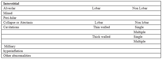
Methods: All the patients came to paediatric department from January 2013 to July 2014 & diagnosed as pneumonia as per WHO protocol were included in study. . It was a prospective, double blind, randomized controlled study of radiological data of all the patients who were diagnosed as pneumonia as per WHO protocol. All the x- rays were read by two experts.
Results: Of 219 radiographs examined 23 have evidences of pneumonia. 183 patients have normal x-ray.
Conclusion: Radiology is consider most accurate method for diagnosis of pneumonia. As per WHO protocol these children receive unnecessary antibiotics although many of these children have viral pneumonia. There is need of some cheaper, easily available & more accurate methods to identify etiological organism for pneumonia.
Downloads
References
2. Under five mortality. Available online at: http://www.who.int/gho/child_health/mortality/mortality_under_five/en/ cited on 22/10/2014
3. Global Health Observatory Data Repository. Aailable online at: http://apps.who.int/gho/data/node.main.38 cited on 22/10/2014
4. Wardlaw T, Salama P, Johansson EW, Mason E. Pneumonia: the leading killer of children. Lancet 2006;368:1048-50. PMID:16997649 doi:10.1016/S0140-6736(06)69334-3
5. Bryce J, Victora CG, Habicht JP, Black RE, Scherpbier RW. Programmatic pathways to child survival: results of a multi-country evaluation of Integrated Management of Childhood Illness. Health Policy Plan 2005;20 Suppl 1;5-17. doi:10.1093/heapol/czi055
6. Dawson P, Pradhan Y, Houston R, Karki S, Poudel D, Hodgins S. From research to national expansion: 20 years’ experience of community-based management of childhood pneumonia in Nepal. Bull World Health Organ 2008;86:339-43
7. World Health Organization. Acute respiratory infections in children: case management in small hospitals in developing countries. WHO/ARI/90, 5th ed. Geneva:WHO, 1990.
8. Campbell H, Byass P, Greenwood BM. Simple clinical signs for diagnosis of acute respiratory infections [letter]. Lancet 1988;2:742-3.
9. Cherian T, John TJ, Simoes E, Steinhoff MC, John M. Evaluation of simple clinical signs for the diagnosis of acute lower respiratory tract infection. Lancet 1988;2:125-8
10. Morley CJ, Thornton AJ, Fowler MA, Cole TJ, Hewson PH. Respiratory rate and severity of illness in babies under 6 months old. Arch Dis Child 1990;65:834-7.
11. World Health Organization. Programme for the control of acute respiratory infections. Report of a meeting of the radiology working group, Geneva 27-28 October 1989. Geneva: WHO,1990, WHO/ARI/90.13.
12. Neuman MI, Lee EY, Bixby S, Diperna S, Hellinger J, et al. (2012) Variability in the interpretation of chest radiographs for the diagnosis of pneumonia in children. J Hosp Med 7: 294–298.
13. Johnson J, Kline JA (2010) Intraobserver and interobserver agreement of the interpretation of pediatric chest radiographs. Emerg Radiol 17: 285–290
14. Stickler GB, Hoffman AD, Taylor WE. Problems in the clinical and roentgenographic diagnosis of pneumonia in young children. Clin Pediatr 1984;23:398-9.
15. Puig S, Hörmann M, Sandström S, Schibany N, Ponhold W. [Acute, atraumatic changes of the lower respiratory tract in the child in thoracic roentgen imaging. Recognition and appreciation of radiological changes. Radiologe. 2002 Mar;42(3):153-61.
16. Bettenay FA1, de Campo JF, McCrossin DB. Differentiating bacterial from viral pneumonias in children. Pediatr Radiol. 1988;18(6):453-4.
17. Tupasi TE, Lucero MG, Magdangal DM, Mangubat NV, Sunico ES, Torres CU, et al. Etiology of acute lower respiratory tract infection in children from Alabang, Metro
Manila. Rev Infect Dis 1990;12:S929-39.
18. Murphy CG1, van de Pol AC, Harper MB, Bachur RG. Clinical predictors of occult pneumonia in the febrile child
Acad Emerg Med. 2007 Mar;14(3):243-9. Epub 2007 Jan 22.
19. Eber E. Treatment of acute viral bronchiolitis. Open Microbiol J. 2011;5:159-64. doi: 10.2174/1874285801105010159. Epub 2011 Dec 30